
Dental Cone Beam CT
To help provide the best possible care for our patients, we recently added a Dental Cone Beam Computed Tomography machine to our office.
Dental Cone Beam Computed Tomography (CBCT) is a special type of x-ray equipment that is used to provide additional information to what is typically obtained from a conventional dental x-rays. We may use this technology to produce three dimensional (3D) images of your teeth, soft tissues, nerve pathways and bone in a single scan. Images obtained with a CBCT allow for more precise diagnosis and treatment planning.
Cone Beam Computed Tomography or CBCT is a variation of the traditional Computed Tomography (CT) used in medical environments. The CBCT system used in our practice captures information using a cone-shaped x-ray beam, which is then used to construct a 3D image of the area of interest.
The CBCT scan captures all the anatomy in one single rotation, subjecting the patient to far less radiation exposure. CBCT offers higher resolution, sharper images, and better artifact reduction compared to medical CT.
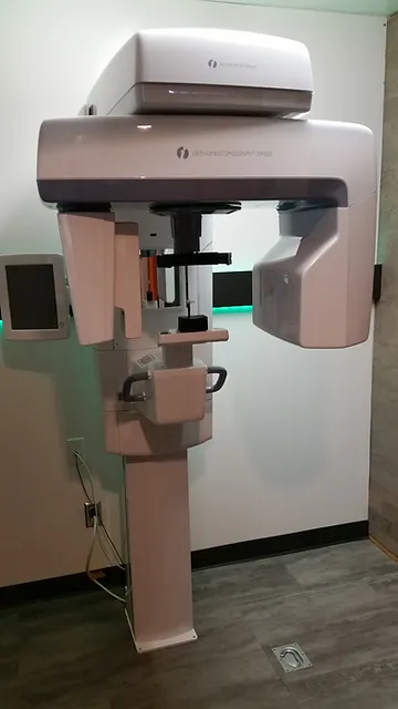
What happens during the CBCT scan?
Prior to the scan, you will be asked to remove any jewelry, piercings, eyeglasses, dentures, hearing aids and hairpins, which would affect the quality of the CT images. Female patients also need to let us know if there is a possibility that they may be pregnant.
During the scan, you will be positioned appropriately in the CBCT machine. The machine has an open design and can accommodate patients of all sizes. A trained member of our staff will carefully position your head and ask you to keep still during the scan. The scan takes less than a minute to complete.
What are the benefits to CBCT scans?
Cone Beam CT provides detailed images of the teeth and bone and is performed to evaluate the status and diseases of the jaws, teeth, bony structures of the face and sinuses. It does not provide the full diagnostic information available with conventional CT, particularly for evaluation of soft tissue structures such as muscles, lymph nodes, glands and nerves. However, cone beam CT has the advantage of far lower radiation exposure compared to conventional CT.
The CBCT is very effective in diagnosing cracks or fractures of the teeth that extend below the gums, bony lesions associated with periodontal disease or infections, amount of bone present for implant placement, impactions, orthodontic evaluation and many other conditions.
The safety of the scan, combined with the quantity of information obtained makes the CBCT an invaluable tool in providing the best possible care for our patients.
What are the risks associated with CBCT?
There is a small amount of radiation exposure with any x-ray or radiologic procedure. Our practice only prescribes CBCT when the situation calls for it. With the advent of CBCT, it is gradually becoming the Standard of Care is some procedures. When we perform a CBCT scan, we adhere to the As Low as Reasonably Achievable (or ALARA) Principle to ensure your complete safety.
CT scanning is in general not recommended for pregnant women, unless medically necessary because of potential risks to the mother or baby.
Because children are more sensitive to radiation, they should only have a CT exam when it is essential for making a diagnosis and with the low-dose technique.
The amount of radiation exposure is comparable to 5 – 7 days of background environmental radiation. Our bodies tolerate low and small amounts of background radiation on a daily basis. The key is to avoid large quantities of radiation in a short span of time, and to prescribe the CBCT when the benefits of the diagnostic data obtained from it outweighs the low radiation exposure. Please let us know if you have any questions or concerns.
Panoramic X-ray Capabilities
Our CBCT machine can also produce a Panoramic Image of the jaws (Panorex) which will be beneficial when assessing erupting or missing teeth in the mixed dentition stage, impacted wisdom teeth, bony lesions, cysts, fractures, bony abnormalities and infections of the jaws.
As with all of our other radiographic services, we can email or send these images to our local Orthodontists, Oral Surgeons or Specialists who may also be providing services and treatment for you.